Saturday, 27 July 2024 09:18 AM
OR
Login using
#Book Review
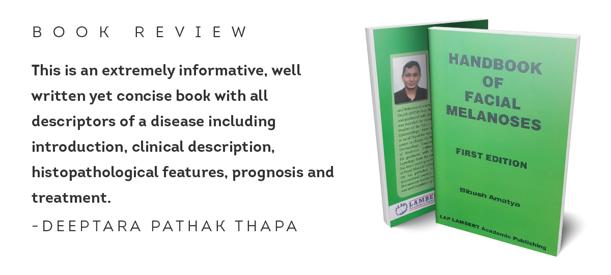
This is an extremely informative, well written yet concise book with all descriptors of a disease including introduction, clinical description, histopathological features, prognosis and treatment. The clinical photographs, Woods lamp photographs, dermoscopic features photographs are of very good quality and are tagged with easily recognizable symbols.
Dr Bibush Amatya’s Handbook of Facial Melanosis, the first edition of Lap Lambert Academic Publishing, is an excellent book worth reading and seems well mounted for postgraduate medical students, young dermatologists and senior consultants alike, who want to improvise their practice with dermoscopy.
The book covers various diseases seen on the face like different types of naevi, common tumors, different types of pigmentation etc. It has been made very interesting, incorporating dermoscopic features, Woods lamp pictures, clinical photos and mentioning a brief introduction of each disease with epidemiology, etiology, the histopathological features along with prognosis and treatment of the diverse group of diseases. The dermoscopic features are coherently written and vividly depicted as different symbols like star, circle, rectangle, square etc. which are self-explanatory and easy for the readers to understand.
In chapter 2, different types of acquired melanocytic naevi are discussed, describing how a junctional nevus can be dermoscopically differentiated from an intradermal nevus or a compound nevus. Characteristic dermoscopic finding of junctional nevus being the reticulated pigment network with central brown dots, grouped bluish gray globules in compound nevus and bluish white structures in intradermal naevus.
In chapter 6, interesting findings like diffuse reddish-brown pigmentation, hem like pigment pattern and reticular pattern are seen in clofazimine induced pigmentation which is very informative.
In chapter 13, various types of melasma are discussed. Dermoscopic features of well-defined networks are seen in epidermal melasma whereas less well-defined networks in dermal melasma. Differentiating melasma with the aid of dermoscopy helps the treating physician for choosing treatment modality for different types of melasma.
In chapter 16, a very uncommon condition like peri-buccal pigmentation of Brocq has been mentioned. The characteristic dermoscopic features are erythema, perifollicular scaling, enlarged follicular opening with yellowish keratotic plugs surrounded by slate blue globules. This adds to the knowledge in the field of dermoscopy.
Tumors, the name itself not only brings psychological stress to the patients but also creates a diagnostic dilemma for dermatologists. Being able to distinguish between benign and malignant tumors dermoscopically, is an important tool which helps decide upon conservative management over surgery and is a boon for treating physicians. Diagnosing benign lesions like seborrheic keratosis has been nicely described in chapter 20 and is very useful. Clinically different variants as well as histopathological variants are mentioned. Characteristic dermoscopic features with histopathological correlation have been nicely put forward which help the readers to understand the features in depth. Dermoscopic features like gyri and sulci histopathologically correlate with pseudo follicular openings on the surface. Other features like milia-like cysts, comedo-like opening, cerebriform pattern have nicely been depicted. Similarly in chapter 21, solar lentigo, a benign pigmented condition, has been described; its dermoscopic features are comedo-like opening, diffuse opaque brown pigmentation, and finger-prints like features are worth mentioning.
Overall this is an extremely informative, well written yet concise book with all descriptors of a disease including introduction, clinical description, histopathological features, prognosis and treatment. The clinical photographs, Woods lamp photographs, dermoscopic features photographs are of very good quality and are tagged with easily recognizable symbols. I personally feel honored to review this book and would highly recommend this book to the readers. A book of such kind is a gift to the field of dermoscopy. (The author is Associate Professor at Nepal Medical College and Teaching Hospital)
You May Like This
#Book Review: The Old Man and The Sea
This book is the last fiction written by the famous writer Ernest Hemingway. It gives us the moral lesson to... Read More...
Aaradhana: A great inspiration for youth
In his school, his teachers from Darjeeling made an indelible mark in his young mind, that English was the superior... Read More...
‘Art Evolves: Nepali Modern Art’: Review
The basic flow of the story goes from introducing the topic and exploring the extent of modernism through a philosophical... Read More...
NRB unveils monetary policy
19 hours ago

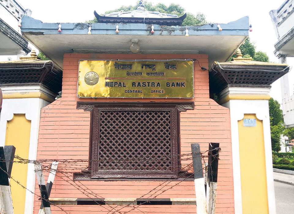
NRB set to issue monetary policy today
21 hours ago
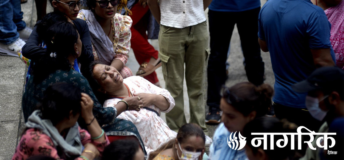
Girl found unconscious at Gwarko guest house dies
22 hours ago
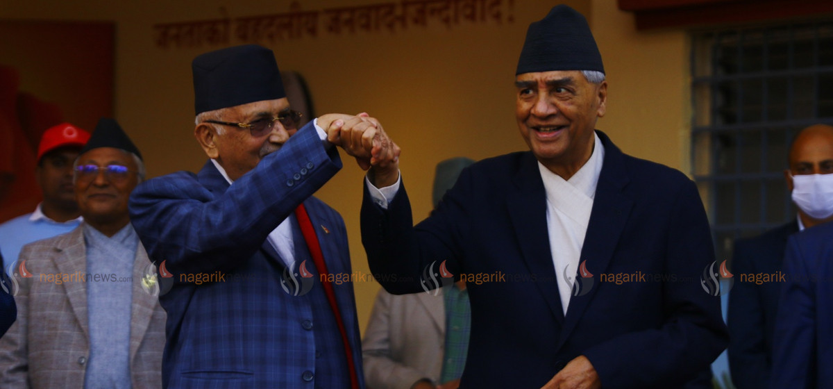
Challenges Confronting the New Coalition
23 minutes ago
NEB to publish Grade 12 results next week
11 hours ago
NC defers its plan to join Koshi govt
13 hours ago
NRB to review microfinance loan interest rate
13 hours ago
Just In
- Challenges Confronting the New Coalition
- NRB introduces cautiously flexible measures to address ongoing slowdown in various economic sectors
- Forced Covid-19 cremations: is it too late for redemption?
- NRB to provide collateral-free loans to foreign employment seekers
- NEB to publish Grade 12 results next week
- Body handover begins; Relatives remain dissatisfied with insurance, compensation amount
- NC defers its plan to join Koshi govt
- NRB to review microfinance loan interest rate
Leave A Comment