Friday, 26 April 2024 12:50 AM
OR
Login using
Autism Appears Unrelated To Ultrasounds During Pregnancy
Published On: February 27, 2018 12:24 PM NPT By: Agencies
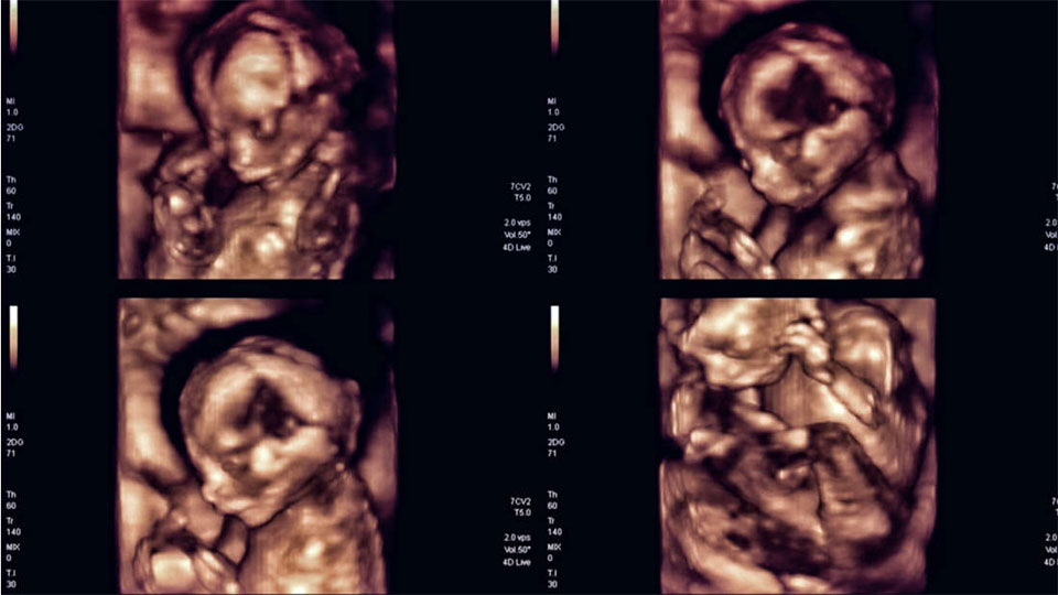
Feb 27: A study led by the Boston University School of Medicine has eased speculative fears after revealing no link between the number or duration of prenatal ultrasounds and a child’s later development of autism spectrum disorder (ASD).
The findings represent one more in a stream of investigations that have failed to identify a direct genetic or environmental cause of ASD, though a variety of factors are now known to increase the risk of development.
In recent years, high-powered ultrasound devices – the kind that can create a 3D picture of a fetus’s features – have become an increasingly popular tool for monitoring pregnancy. Because rates of ASD diagnosis have risen sharply during the same time frame, fetal exposure to ultrasound was quickly implicated as a potential factor for its cause. Some subsequent experiments in animal fetuses suggested that these devices can modify brain formation, leading to changes in memory, learning, and sociability once the organism is born.
To evaluate the link in humans, the authors of the current investigation, published in JAMA Pediatrics, reviewed the medical records of 107 otherwise healthy children with ASD, 104 children with a non-ASD developmental delay, and 209 children with normal neurological development.
The data showed that normal children and those with ASD or developmental delays were all exposed to the same average number of ultrasounds (about six) as fetuses, and that the time taken for each scan was similar.The scans’ average thermal index – the approximated amount of fetal tissue warming that occurs during a scan – were also the same between groups.
“In almost every parameter we looked at, ultrasound seemed perfectly safe,” co-author N. Paul Rosman toldThe Washington Post.
The only difference between groups was the depth that ultrasound waves permeated into the maternal and fetal tissue. Scans of ASD fetuses had a slightly greater average depth during the first and second trimesters compared with scans performed on normal fetuses.
Ultrasonography consists of sending pulses of high-frequency sound into tissue and creating an image from the pattern of reflected or scattered soundwaves. Though the frequency used for medical imaging has long been considered safe, the FDA and the American College of Obstetricians agree that exposure should be limited due to the possibility of undiscovered side effects.
Thus, the authors noted that the deeper scan depth could lead to increased penetration of soundwaves through the skull and into the fetal brain, potentially altering the brain cell development occurring within. They conclude by calling for future studies into ultrasound scan depth and ASD.
However, other pediatric specialists, like the two physicians who wrote an accompanying editorial, are skeptical that such a small difference in scan depth could account for the development of a severe neurological condition such as ASD.
You May Like This
NHRC to monitor human rights situation during local polls
KATHMANDU, April 27: National Human Rights Commission (NHRC) said that the constitutional body will monitor human rights situation across the... Read More...

Traffic not to be halted during Indian President's visit
KATHMANDU, Oct 30: Efforts are on to making the vehicular traffic smooth and hassle-free for the general public during the... Read More...
2,870 structures damaged during Maoist conflict reconstructed: Ministry
KATHMANDU, July 19: The total 2,870 structures destroyed during the Maoist decade-long insurgency have been reconstructed until 15 July 2016,... Read More...


Karnali CM Kandel secures vote of confidence
7 hours ago
Just In
- World Malaria Day: Foreign returnees more susceptible to the vector-borne disease
- MoEST seeks EC’s help in identifying teachers linked to political parties
- 70 community and national forests affected by fire in Parbat till Wednesday
- NEPSE loses 3.24 points, while daily turnover inclines to Rs 2.36 billion
- Pak Embassy awards scholarships to 180 Nepali students
- President Paudel approves mobilization of army personnel for by-elections security
- Bhajang and Ilam by-elections: 69 polling stations classified as ‘highly sensitive’
- Karnali CM Kandel secures vote of confidence
Leave A Comment